Endocardium inner layer consists of endothelium connective tissue. The e ndocardium myocardium and e picardium.

Pin On Human A P
Terms in this set 3 Epicardium outer layer consists of epithelium connective tissue.

Heart 3 layers. The innermost layer of the cardiac wall is known as the endocardium. The inner layer of. Epicardium myocardium and endocardium.
The heart wall consists of three layers Fig. The color of the heart should be a deep rich brown. The Layers of the Heart Wall The heart wall is composed of connective tissue endothelium and cardiac muscle.
Layers of the Heart Wall. Epicardium myocardium and endocardium. It is the cardiac muscle that enables the heart to contract and allows for the synchronization of the heartbeat.
A beautiful 3 layered beaded heart available in Clear Silver or White beads. What are the three layers called. Large measures 38x38cm Measures 28x28cm Small 16x16cm.
The outermost layer of the wall of the heart is also the innermost layer of the pericardium the epicardium or the visceral pericardium discussed earlier. This layer covers the outer layer of the heart and consists of a single layer of mesothelial cells and connective tissue. Myocardium middle layer cardiac muscle layer.
The heart wall is comprised of three layers the epicardium outer myocardium middle and endocardium inner. Three layers of the heart wall. The heart wall is divided into three layers.
It helps to keep the heart lubricated and protected. The outer protective layer of the heart. Three layers of the heart.
Like other vessels our heart is composed of three distinct layers. There are three layers that make up the heart muscle. The heart wall itself can be divided into three distinct layers.
The surface layer of the heart epicardium is directly below the fibrous and parietal pericardium. The epicardium is also sometimes considered a part of the protective pericardial membrane around the heart. Epicardium Myocardium and the Endocardium.
These sulci run along lines that separate atria from ventricles atrioventricular sulcus as well as right and left sides of ventricles interventricular sulcus. Beneath the body of the espresso shot is the heart of the espresso at the bottom of the shot. Muscular middle layer wall of the heart.
Hand decorated and hang from diaphanous ribbon. It lines the cavities and valves of the heart. The first layer is the epicardium which is the visceral layer of the serous pericardium.
Structure of the Heart. From superficial to deep these are the epicardium the myocardium and the endocardium. Giving a lovely effect.
Each of these layers have a different role for heart contraction. The heart has three layers of tissue each of which serve a slightly different purpose. Histologically the heart is made of three layers of tissue.
Heart Exterior. Within the heart of the espresso shot are the shots bitter qualities which balance out the. 2 a b and c.
In this article we are going to dissect each layer and to understand the pathology that can happen within each of these layers. Layers of the Heart Wall. In this article we shall look at the anatomy and clinical relevance of these layers.
The external heart surface contains grooves or sulci which provide passageways for blood vessels of the heart. With pictures - wiseGEEK What are the three layers of heart tissue Sat May 11 2019 The Layers of the Heart Wall. The wall of the heart is composed of three layers of unequal thickness.
The epicardium the myocardium and the endocardium. The human heart is a four-chambered muscular organ shaped and sized roughly like a mans closed fist with two-thirds of the mass to the left of midline. The heart is enclosed in a pericardial sac that is lined with the parietal layers of a serous membraneThe visceral layer of the serous membrane forms the epicardium.
These tissue layers are highly specialized and perform different functions. Arteries and Veins lay travel through this layer and goes deep in the hearts layers to feed it Oxygen from the arteries and veins which carry Deoxygenated blood back into the R Atrium. The Epicardium is the surface of the heart.
Has three different hearts which move independently.

Medical Careers Pearson Education Muscle Tissue
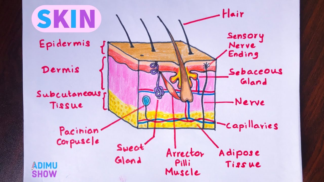
How To Draw Skin Layers Integumentary System Step By Step Drawing Youtube Integumentary System Medical School Inspiration Nursing School Notes

Layers Of The Pericardium Heart Wall And Spiral Arrangement Cardiovascular System Biology Lessons Medical Anatomy

Atriums And Layers Of The Heart Atrium Layers Inspiration

Coronary Heart Layers Pericardium Endocardium Myocardium Anatomy Physiology Cardiovasc Human Anatomy And Physiology Heart Anatomy Medical Assistant Student

Cardiac Anatomy Creative Commons Illustration Radiology Case Radiopaedia Org Human Anatomy Picture Cardiac Anatomy Human Heart Anatomy

Cardiovascular System Powerpoint And Notes Distance Learning Cardiovascular System Cardiovascular High School Science Teacher

Cardiovascular Structures And Layers Of The Heart Youtube Cardiovascular Science Layers

19 6 Pericardium The Protective Layers Of The Heart Include The Pericardial Sac Composed Of An Outer Anatomy Models Human Anatomy And Physiology Heart Anatomy

Heart Wall And Its Parts Heart Structure Heart Function Human Heart Diagram

How To Draw Internal Structure Of Human Heart Easy Version Heart Diagram Human Heart Diagram Easy Heart Drawings
Picture 3 Layers Diastasis Recti Diastasis Rectus Muscle

Pin On College

Love In Layers Candy Heart Cake Wilton Cake Decorating Cupcake Cakes Cake Topper Tutorial

Pin On A P Heart

The Heart Wall Is Made Up Of 3 Layers That Have Their Own Functions Heart Wall Human Heart Diagram Heart Anatomy

Pericardium Serous Membranes Visceral Parietal Layers Medical Coding Human Anatomy And Physiology Serous Membrane

Layers Of The Heart Nursing School Nurse Nurturing

Pin On Biology 100

